Da Wang
Ornstein Laboratory, room 0.14
Princetonplein 1, 3584 CC Utrecht
P.O. Box 80 000, 3508 TA Utrecht
The Netherlands
phone: +31(0)30 253 1287
secretariat: +31(0)30 253 2952
e-mail: d.wang@uu.nl
Research
Promotor: Prof. dr. Alfons van Blaaderen
Funding: EU-ERC Advanced Grant
Employed: 1 August 2012 – 31 October 2018
Self-assembled Supraparticles from nanocrystals
Colloidal supraparticles which are assembled from size- and morphology- controlled nanoparticles, combine multi-scale properties of the single particles such as quantum confinement and localized plasmon resonances with collective effects resulting from being arranged on a 3D lattice. In addition properties on longer length scales, e.g. photonic, can be controlled by a subsequent self-assembly step. One way to realize such colloidal supraparticles is by suspending nanocrystals in emulsion droplets of a low boiling point solvent in water and slowly evaporating the solvent. In this way the nanocrystals are forced to self-assemble into supraparticles. By tuning the concentrations and types of nanocrystals, supraparticles with different structures and sizes can be obtained.
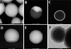
The goal of this research is to extend the spherical confinement method to binary particles systems and anisotropic particles systems. For instance, 7.6 nm CdSe nanocrystals and 9.9 nm PbSe nanocrystals were used to synthesize binary supraparticles with a bulk MgZn2 phase. We found 16.5 nm Ag/9.1 nm Fe3O4 and 6.5 nm PbSe/9.3 nm Au self-assembled into Janus shape binary supraparticles or core-shell binary supraparticles, respectively, which probably can be ascribed to attractive interactions between different ligands. EuF3 round platelets nanocrystals can also self-assemble into supraparticles with liquid-crystal-like interior structure. In addition, supraparticles were successfully assembled by using rounded FexO/CoFe2O4 nanocubes as building blocks. After freeze drying, the structure of the supraparticles was studied with high-angle annular dark-field scanning transmission electron microscopy (HAADF-STEM) tomography and secondary electron scanning transmission electron microscopy (SE-STEM). By coating the supraparticles with a thin (~50 nm) layer of porous or non-porous silica, the particles become more robust and do not deform by drying on a substrate. To study how the structure of the more complex supraparticles is affected by the spherical confinement, work is in progress by advanced electron microscopy, 3D particle fitting technique and computer simulations.
Figure 1: A) HAADF-STEM image of PbSe/CdSe binary supraparticles; B) HAADF-STEM image of Ag/Fe3O4 Janus supraparticles; C) SE-STEM image of PbSe/Au core-shell supraparticles; D) HAADF-STEM image EuF3 round platelets supraparticles; E) HAADF-STEM image round edge cube FexO/CoFe2O4 supraparticles; F) TEM image of the Au supraparticles coated with mesoporous silica shell. (Supraparticles in Fig A, C, D: collaboration with the C. B. Murray group (U. Pennsylvania, USA))

