
Ernest van der Wee
Leonard S. Ornstein Laboratory, room 0.14
Princetonplein 1, 3584 CC Utrecht
P.O. Box 80 000, 3508 TA Utrecht
The Netherlands
phone: +31 (0)30 253 1287
secretariat: +31 (0)30 253 2952
e-mail: e.b.vanderwee@uu.nl
Research
Promotor: Prof. dr. Alfons van Blaaderen
Funding: EU-ERC Advanced Grant
Employed: 15 October 2013 – 1 July 2018
Arresting colloidal self-assemblies and processes
Confocal laser fluorescence microscopy (CLSM) is successfully used to acquire 3D information of densely packed colloidal systems [1]. This is, however, much harder for systems with more mobile particles [2]: the particles might move during a single scan, resulting in incorrect 3D information (see figure 1a). This effect can be minimized by scanning faster and increasing the viscosity of the dispersion. In this project, we investigate complete arrest of the particles by polymerizing a component of the solvent. By adding a monomer and initiator to a dispersion of sterically stabilized poly(methyl methacrylate) (PMMA) particles with a Yukawa potential [2] and exposing them to UV light, we arrest the crystal. Such a procedure was developed for aqueous systems by, amongst others, the Asher group [3]. We extract 3D coordinates from the CLSM images prior to, and after, the polymerization (see figure 1b). In addition, we expose the particles to UV light while scanning the sample, to gain insight on the timescale of the arrest. We study quantitatively, if, and how, the configuration of the system changes during polymerization.
This method can be used to study colloidal self‐assemblies and processes on a fundamental level (e.g. structure and nucleation of crystals), as well as for interesting applications. For instance: the exposure of polymerized colloidal crystals to electric fields, yielding tunable photonic crystals.
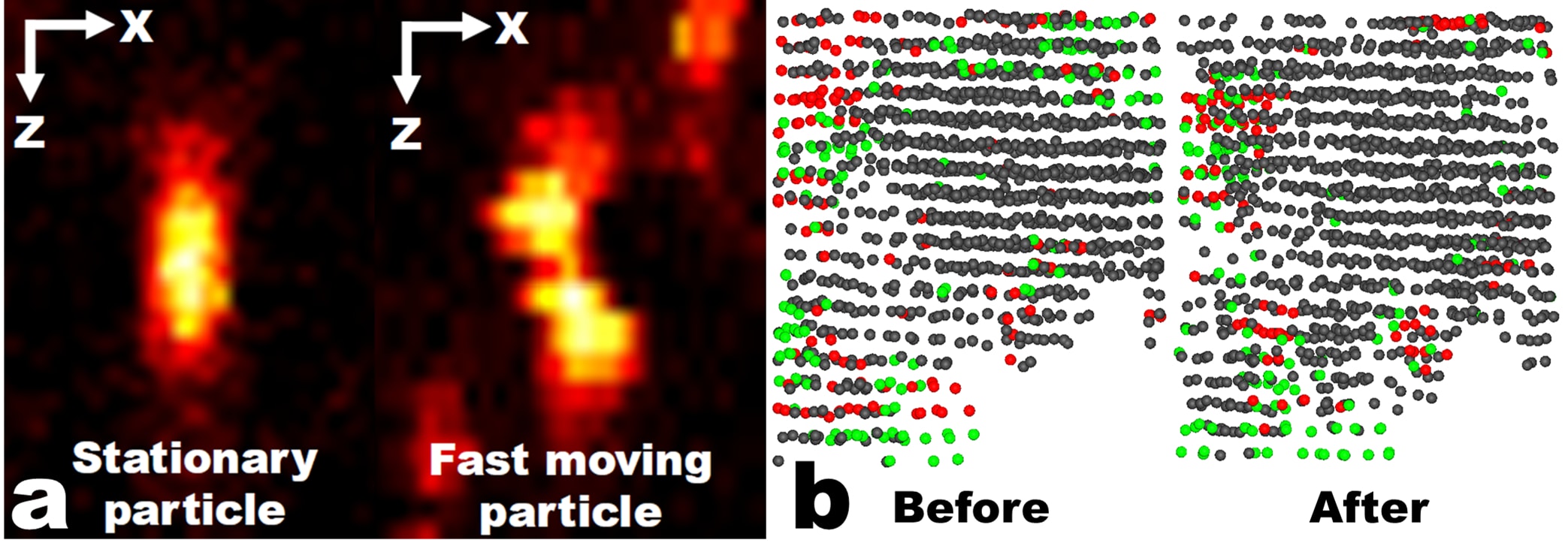
Figure 1: a) Confocal images in the XZ plane of a stationary and a fast moving colloid. b) Computer reconstruction of the coordinates of the colloidal particles before and after the arrest. Only crystalline particles are shown. The colors denote the crystalline order of the particles (red – FCC, green – HCP, gray – BCC).
[1] A. van Blaaderen and P. Wiltzius, Science 270, 1177-1179 (1995)
[2] A. Yethiraj and A. van Blaaderen, Nature 421, 513 (2003)
[3] J.H. Holtz and S.A. Asher, Nature 389, 829-832 (1997)