
Sina Sadighikia MSc
Leonard S. Ornstein Laboratory, room 0.06
Princetonplein 1, 3584 CC Utrecht
P.O. Box 80 000, 3508 TA Utrecht
The Netherlands
phone: +31 (0)30 253 2830
secretariat: +31 (0)30 253 2952
e-mail: s.sadighikia@uu.nl
Research
Supervisor: Dr. Marijn van Huis
Promotor: Prof.dr. Alfons van Blaaderen
Funding: STW
Employed: 1 February 2016 – 31 January 2020
Understanding silica nanoparticles assemblies in solution using In-Situ TEM
Suspensions of colloidal nanoparticles can self-assemble into highly ordered 3D nanostructures called colloidal crystals. Colloidal crystals have attracted significant attention since they are ideal model for understanding the fundamental characteristics of crystallization, melting and phase transformation of materials.
My research focuses on understanding silica nanoparticle assemblies in solution using In-Situ TEM. As a first step, colloidal silica nanoparticles have been synthesized using several methods. Among these methods, highly monodisperse particles can be prepared using a new amino acid catalysed method1. This procedure starts with the nucleation of a silica seed of around 16 nm (Figure 1a) followed with a seeded growth method to produce a desired particle size (Figure 1b). Growing colloidal crystals from these particles is carried out in highly deionized water in presence of ion-exchange resin beads. Colloidal crystals will be examined using Liquid-Cell Transmission Electron Microscopy (LCTEM) (Figure 1c) and Confocal microscopy. In later work, gold particles coated with labelled silica shells will be synthesized. Gold cores are beneficial because they will allow us to work with lower electron doses (silica is a beam sensitive material) giving better contrast in TEM imaging. Furthermore, by producing labelled silica shells we allow ourselves to study the crystals with confocal microscopy using fluorescence techniques.
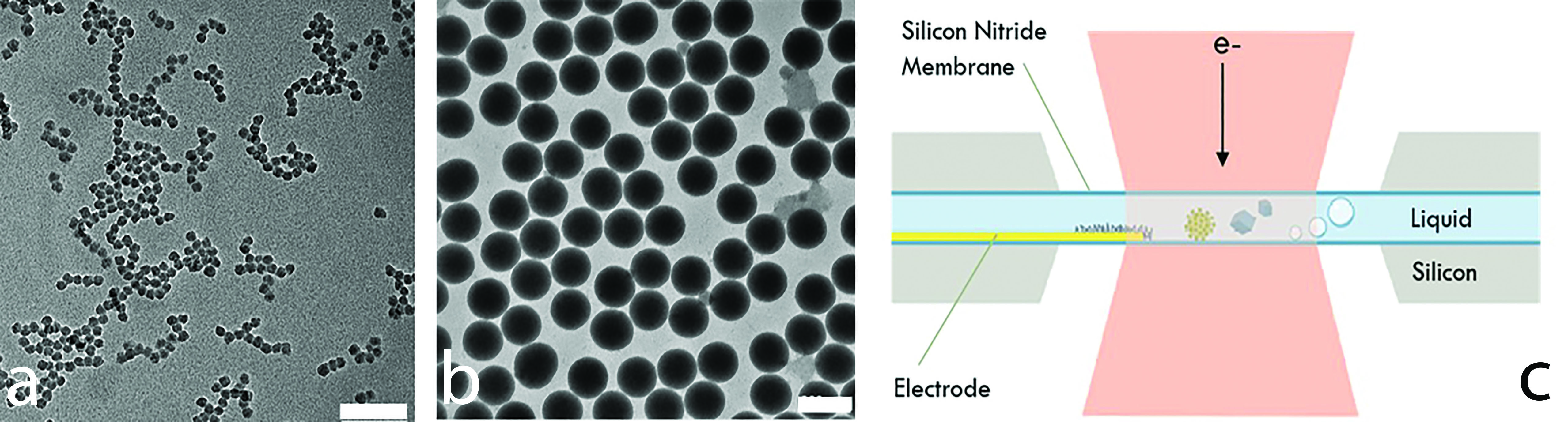
Figure 1: a) Silica seeds with measured diameter of 16 nm and polydispersity of 5% (scale bar is 100 nm). b) Silica nanoparticles with measured diameter of 150 nm and polydispersity of 1% (scale bar is 200 nm). c) Schematic diagram of a liquid cell for the transmission electron microscope
[1] Shahabi et al., J. Nanoparticle Res. 17, 270 (2015).