Tom Welling MSc
Ornstein Laboratory, room 0.06
Princetonplein 1, 3584 CC Utrecht
P.O. Box 80 000, 3508 TA Utrecht
The Netherlands
phone: 030 253 2830
secretariat: +31(0)30 253 2952
e-mail: t.a.j.welling@uu.nl
Research
Supervisor: Dr. Marijn van Huis
Promotor: Prof. dr. Alfons van Blaaderen
Funding: ERC
Employed: 15 February 2017 – 14 February 2021
Self-assembly of nanoparticles in liquid cell electron microscopy
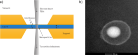
Liquid cell (scanning) transmission electron microscopy (LC-(S)TEM) is a promising new technique to acquire information on nanoparticle movement and positions with high spatial resolution [1]. By employing a cell with electron transparent windows that separates a thin liquid layer (< 5 micron) from the vacuum of the microscope (Figure 1a), it is possible to image nanoparticles with nm resolution in a liquid environment.
While the interactions between micron-sized colloids are relatively well understood, many assumptions break down at the nanoparticle size regime (< 20 nm) [2]. The high spatial resolution of electron microscopy can resolve small particles and their position relative to each other. This allows us to monitor the interactions between particles and their assembly in situ.
Interesting and convenient systems to study are so-called rattler particles [3], which consist of a core particle which can move in a void enclosed by a shell (Figure 1b). It is possible to track the cores within the shell, which is the first step to obtaining meaningful data from liquid cell experiments. We can use tracking data to probe the interactions between the core and the shell and learn about the challenges of this new technique, such as electron-solvent interactions.
At a later date, we will attempt to monitor the assembly of nanoparticles in situ by making use of solvent-evaporation, heating or electric fields inside the electron microscope.
Figure 1: a) Geometry of a liquid cell [1]. b) Rattler particle consisting of a titania core and silica shell [3] imaged in liquid in an electron microscope.
[1] De Jonge and Ross, Nature Nanotechnol. 6, 695-704 (2011)
[2] Silvera Batista et al., Science 350, 1242477 (2015)
[3] Watanabe et al., Langmuir 33, 296-302 (2017)
