
Henriëtte Bakker
Dr. H.E. Bakker
Research
Supervisor: dr. A. Imhof
Promotor: Prof.dr. A. van Blaaderen
Employed since April 2012
Funded by FOM
Colloids, Confocal Microscopy, Particle Tracking
Charged particle dispersions, with particles ranging from the nano- to micrometer range, are present in everyday life. One could think of small DNA strands, proteins, fd-virus, micelles or charged colloids, such as present in e-ink, printer toners. Therefore, investigating their behavior in an electric field of these charged particles is of substantial importance in biophysics and industry. Most of these particles have a rod-like shape. Silica rods are short and rigid, this makes them ideal, to use as model particles in electrophoresis studies.
Moreover, these dielectric silica rods can also be used to study the out of equilibrium behaviour of rigid rods driven by external fields. Interactions between the rods can be modified by changing the Debye double layer thickness κ-1, which can be done by changing the ionic strength of the medium or changing the medium in which the particles are dispersed. Studying the properties of a single rod under the influence of an external field is of importance to understand the collective behavior in more dense systems.
We study the movement of silica rods during electrophoresis using confocal microscopy. Most measurements on the electrophoresis on rods are done using light scattering techniques. On the other hand, using confocal microscopy and particle tracking algorithms, one can measure the electrophoretic mobility of rod-like particles one the single particle level [1]. Using these techniques we investigate and quantify the movement of the silica particles under the influence of an electric field, and as function of their double layer thickness κ-1.
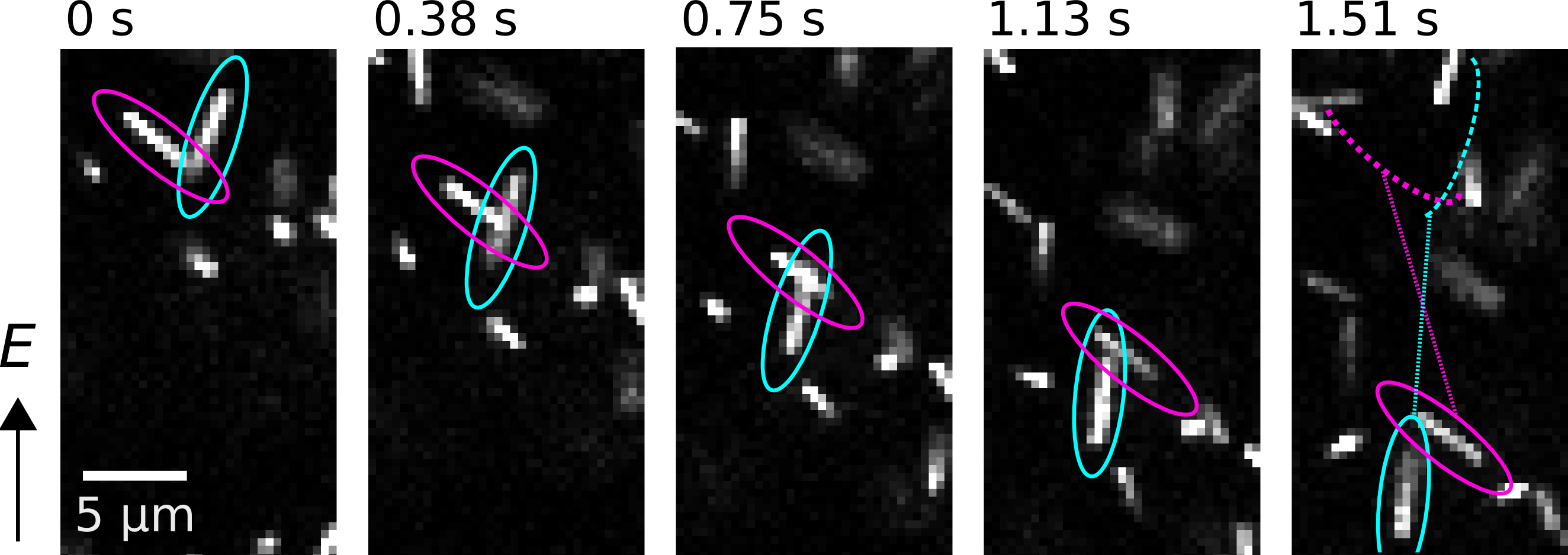
Figure 1: Confocal images of xyt-series of the electrophoresis of silica rods in DMSO-water. Silica rods show an angle dependent velocity and motion at an angle to the applied electric field.
[1] M.G.L. van den Heuvel et al., Proc. Natl. Acad. Sci. 104, 7770–7775 (2007).
Colloidal synthesis, Confocal laser scanning microscopy (CLSM), Particle tracking, Scanning electron microscopy (SEM)
Colloidal silica rods can form, due to their anisotropy, not only a gas, liquid and crystal phase but also liquid crystalline phases. In these phases the rods possess, next to positional order, orientational order. When the silica rods are mixed with silica spheres and left to sediment an intermediate phase is observed in where the spheres fill up the space in between the smectic rods of rods. This binary smectic phase has previously been called a lamellar phase [1,2].
Using fluorescently labeled silica particles and dispersing them in an index-matched medium we can study the phase behavior using confocal scanning laser microscopy (CLSM). With the aid of particle identification algorithms [3,4,5] we study in real space and in solution the structure of the binary smectic liquid crystalline phase. In this way the particle density as function of height can be determined, and therefore the equation of state of the binary mixture can be obtained. By changing the initial composition and volume fraction of the samples, we try to map out the phase diagram for this system. These results indicate that spherical colloids can be incorporated in a crystal of rods, which may have interesting photonic properties.
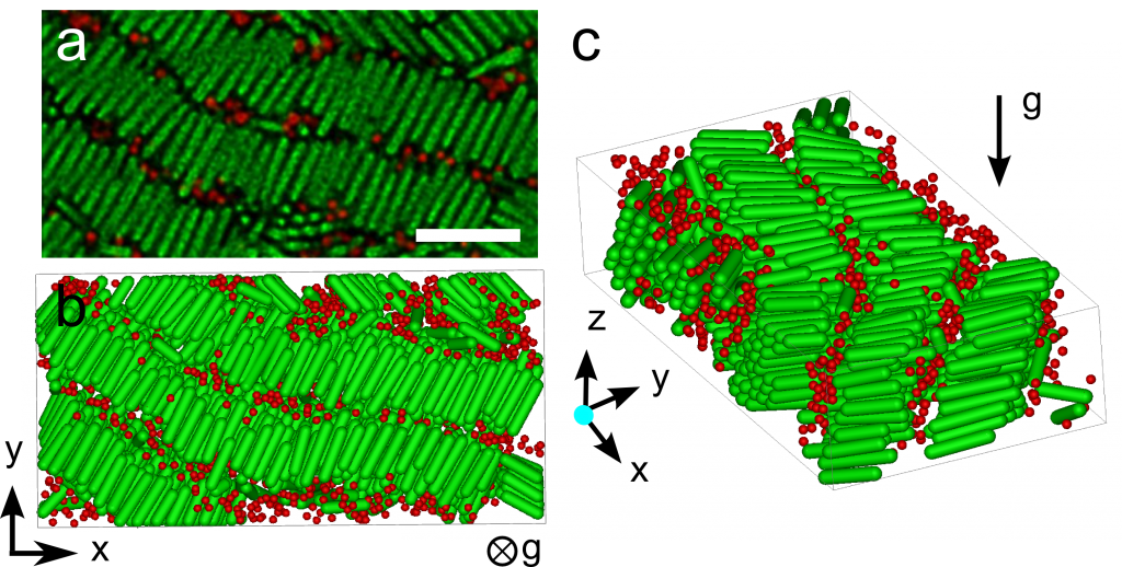
Figure 1 (a) confocal image of a binary smectic phase (b-c) 3D computer reconstruction of a binary smectic phase obtained from experimental confocal data. (b) view in xy-plane (c) xyz-view.
[1] Adams et al., Nature 393, 349 (1998)
[2] Ye et al., Nano Lett. 13, 4980 (2013)
[3] Besseling et al., accepted in J. Phys.: Condens. Matter
[4] Crocker and Grier, J. Colloidal Interface Sci. 179, 298 (1996)
[5] Dassanayake et al., J. Chem. Phys. 112, 3851 (2000)